Benign skin tumours: when removal is needed
What are benign skin tumours?
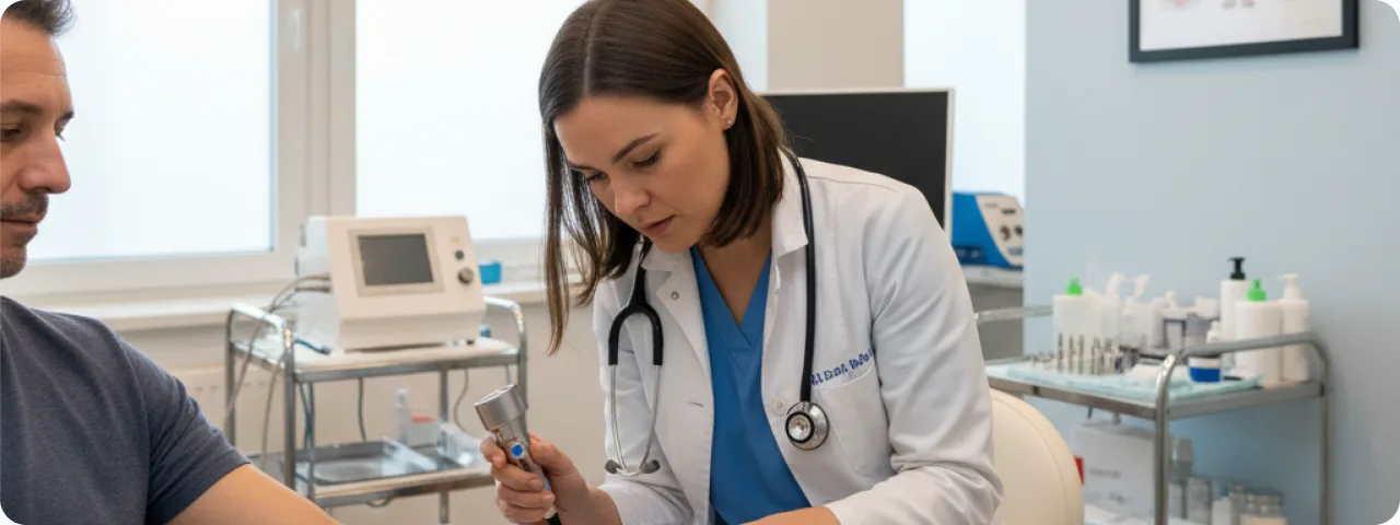
Benign skin tumours are growths that arise from excessive multiplication of the cells of the various layers of the skin, but have no capacity for invasive growth or metastasis. The most common are: papillomas (soft growths on a stalk, caused by human papillomavirus), seborrhoeic keratoses (age warts with a characteristic waxy surface), fibromas (firm nodules of connective tissue), lipomas (subcutaneous growths of fatty tissue), haemangiomas (vascular lesions of a bright red or bluish colour) and dermatofibromas. Statistically, up to 80% of people over 40 have at least one benign skin lesion. Most pose no threat to health and need only to be monitored. Some, however, may cause cosmetic concern, be injured by clothing or jewellery, or — rarely — have the potential to become malignant. That is why it is important to understand the criteria that determine whether removal is needed.
Types of benign lesion and their features
Each type of benign tumour has its own characteristics, outlook and approach to treatment. These are the main groups that patients bring to a dermatologist:
- Papillomas and skin tags: soft skin growths on a thin stalk, usually appearing where there is friction — on the neck, in the armpits and groin, and under the breasts. They range from 1 mm to 1–2 cm. They rarely become malignant, but they are often caught by clothing and jewellery, which causes inflammation and bleeding. Removal is indicated where they are repeatedly injured or cause cosmetic concern.
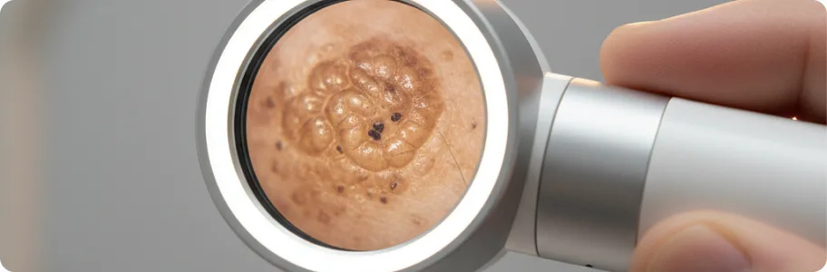
- Seborrhoeic keratoses: firm brown or dark brown plaques with a characteristic warty surface, appearing stuck onto the skin. They usually develop after the age of 40 on the trunk, face and limbs. They are entirely benign, but sometimes need to be distinguished from melanoma — particularly heavily pigmented or rapidly growing ones.
- Dermatofibromas: firm nodules 0.5–2 cm across, most often on the limbs, with a characteristic dimple sign on pressure. They are usually painless and need no treatment. They are removed only at the patient's request, or where their position causes constant injury (for example, an area that is shaved).
- Lipomas: soft, mobile subcutaneous nodules of fatty tissue that grow slowly and rarely exceed 5 cm. They do not become malignant, but when large they can compress nerves or vessels. Indications for removal are rapid growth (which requires liposarcoma to be excluded), pain, a cosmetic defect or restricted movement.
Indications for removal: when waiting is dangerous
Absolute indications for removing a benign lesion include: suspicion of malignancy (rapid growth, a change in colour, shape or surface, bleeding without injury); chronic injury (a position subject to friction from clothing, a belt or jewellery); functional problems (restricted movement, compression of nerves or vessels by a large lipoma); and inflammatory complications (an infected epidermal cyst, recurrent inflammation). Relative indications are primarily the patient's cosmetic concern (particularly on the face, neck and exposed areas), psychological discomfort, and the patient's wish to have the nature of the lesion confirmed histologically. A separate group is lesions with pre-malignant potential: actinic keratoses (a 5–20% risk of progressing to squamous cell carcinoma), dysplastic naevi and Bowen's disease. These must be removed and examined histologically. The decision is always made individually after a dermatological examination and dermatoscopy.
Current methods of removal: from laser to radiofrequency
The choice of method depends on the type, size and site of the lesion and on whether histology is needed. Surgical excision is the classic method and provides material for histology; it is indicated where malignancy is suspected and for large lesions (lipomas over 3 cm). Sutures are removed after 7–14 days, and the scar matures over 6–12 months. Laser removal (CO₂ or erbium laser) provides precise layer-by-layer vaporisation of tissue with minimal injury to the surrounding structures. It is ideal for papillomas, keratoses, small fibromas and superficial lesions on the face. Healing takes 7–14 days and the risk of scarring is minimal. The radiofrequency method (Surgitron) uses high-frequency radio waves to cut without contact and without pressure on the tissue. The zone of thermal damage is minimal and material can still be obtained for histology. Cryosurgery (liquid nitrogen, –196°C) is used for seborrhoeic keratoses, viral warts and actinic keratoses. Its drawbacks are that histological confirmation is impossible and there is a risk of hypopigmentation. Electrocoagulation is effective for small papillomas and vascular lesions.
Recovery and aftercare
Proper care after the procedure is the key to quick healing and minimal scarring. The first 24–48 hours: keep the wound dry, do not use make-up over the treated area, and take pain relief if needed. During the first week: clean the wound with an antiseptic (chlorhexidine or miramistin) twice a day, apply a healing preparation (dexpanthenol or products with hyaluronic acid), and protect it from direct sun. For 2–4 weeks: avoid saunas, swimming pools and open water; do not pick off the crust that forms — it will come away on its own; use SPF 50+ sunscreen on the area once it has healed completely. To prevent hypertrophic and keloid scars, the doctor may recommend silicone sheets or silicone gel (from week 2–3). A follow-up appointment is usually arranged 7–10 days after the procedure. If purulent discharge, marked redness, swelling or fever appear, see a doctor immediately.
When a dermatological consultation is urgent
See a specialist immediately if: the lesion has grown sharply over the past 2–3 months; its colour has changed — becoming darker or unevenly coloured, or developing red and black flecks; the lesion has started to bleed for no apparent reason, or has not healed within 3 weeks of an injury; pain, itching or burning has appeared in the area; or satellite lesions have appeared around the main one. It is also worth seeking advice if you have multiple lesions appearing at the same time or in waves — this can be a sign of systemic disease (for example, the Leser–Trélat sign in internal malignancy). Patients who are immunosuppressed, who have had an organ transplant or who are on long-term immunosuppressive therapy should have a dermatological examination every 3–6 months, since the risk of malignant transformation in this group is considerably higher. Remember: an early consultation does not commit you to removal, but it gives you certainty and a clear plan.
Related reading


Did not find the answer to your question?
Every case is individual. If you have noticed changes in your skin, or have symptoms that concern you, book a consultation.