Melanoma: how to tell a dangerous mole apart
What is melanoma and why is it dangerous?
Melanoma is a malignant tumour of the skin that develops from pigment cells (melanocytes). Although melanoma accounts for only 4% of all cases of skin cancer, it is responsible for 80% of deaths from cutaneous malignancy. More than 325 000 new cases of melanoma are diagnosed worldwide every year. In Ukraine the incidence is rising by 3–5% annually. The main danger of melanoma is its ability to metastasise quickly: a tumour only 1 mm thick can already spread to distant sites through the blood and the lymphatics. When detected early (stage I), five-year survival reaches 98%, whereas at the late stages (IV) it falls to 15–20%. That is why being able to tell a dangerous mole from an ordinary one can literally save a life. Melanoma can arise either from an existing mole (naevus) or on previously clear skin — roughly 70% of melanomas develop de novo and 30% from pre-existing naevi. This means that monitoring must cover not only existing moles but also the appearance of new pigmented lesions.
The ABCDE rule: recognising a dangerous mole
Dermatologists have developed the ABCDE system — five key features that help to distinguish melanoma from an ordinary mole:
- A — Asymmetry: an ordinary mole is symmetrical — if you divide it mentally with a line, the two halves will look the same. Melanoma, by contrast, is asymmetrical: one half does not match the other in shape, size or colour. Check every suspicious mole by dividing it with imaginary lines, horizontal and vertical. If the halves differ substantially in even one direction, that is a reason to see a doctor.
- B — Border: a benign mole has clear, even, smooth edges. Melanoma is characterised by uneven, notched, blurred or ragged edges. The boundary between the pigmented lesion and the surrounding skin is indistinct, and the pigment may spread beyond the main body of the lesion. Pay particular attention to moles whose edges have changed recently.
- C — Color: an ordinary mole has a uniform colour, usually a single shade of brown. Melanoma has uneven colouring: several shades of brown, black, red, white, blue or grey may be combined within one lesion. The appearance of black, blue or white areas within a mole is a warning sign requiring immediate dermatological consultation.
- D — Diameter: moles larger than 6 mm (the size of a pencil eraser) deserve closer attention. Melanoma can be smaller, but statistically larger lesions carry a higher risk of malignancy. E — Evolution: the most important criterion. Any change in a mole over time — growth, a change in shape or colour, the appearance of itching or bleeding, or crusting — is grounds for seeing a dermatologist at once. Photograph suspicious moles once a month for comparison.
Risk factors for melanoma
The risk of melanoma is determined by a combination of genetic and external factors. Ultraviolet radiation is the main modifiable risk factor: intense intermittent exposure (sunburn) is more dangerous than chronic moderate sun exposure. A single severe sunburn with blistering in childhood or adolescence doubles the risk of melanoma in adult life. Using sunbeds increases the risk by 75% where first exposure is before the age of 35. Fitzpatrick skin phototype I–II (fair skin, red or fair hair, freckles, a tendency to burn) is associated with a 2–3-fold increase in risk. The number of moles is one of the strongest predictors: having more than 100 ordinary naevi or more than 5 atypical (dysplastic) naevi increases the risk 6–12 times. Heredity plays a substantial part: 5–10% of melanomas are familial, often linked to mutations in the CDKN2A and CDK4 genes. If a first-degree relative has been diagnosed with melanoma, your risk is 2–3 times higher. Immunosuppression — transplant recipients, patients with HIV and those on immunosuppressive treatment have a 2–8 times higher risk. Previous skin cancer of any type also increases the likelihood of developing melanoma.
Modern diagnosis of melanoma
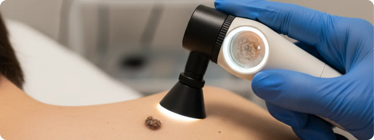
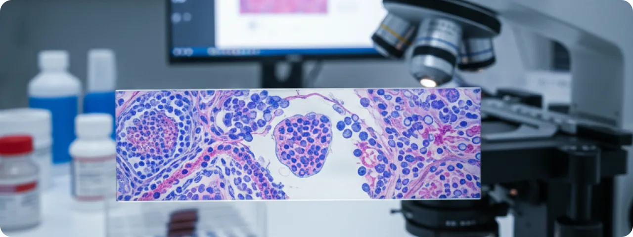
Timely diagnosis is the key to successful treatment. Self-examination of the skin should be carried out monthly, systematically checking the whole body including the scalp, between the fingers and toes, the soles of the feet and the nail plates. Use a mirror for areas that are hard to see, or ask someone close to you to help. Clinical examination by a dermatologist uses the ABCDE rule and the "ugly duckling sign" — a mole that looks different from all the others on the body deserves particular attention. Dermatoscopy is the key non-invasive diagnostic method and raises the accuracy of recognising melanoma from 65–75% (clinical examination) to 90–95%. The dermatoscope makes it possible to visualise skin structures invisible to the naked eye: the pigment network, radial streaming, the blue-white veil and atypical vascular patterns. Digital dermatoscopy with total body mapping is the gold standard for monitoring high-risk patients: every mole is photographed and compared with previous images after 3–6 months. Confocal laser microscopy allows the cellular structure of the skin to be visualised in vivo, reducing the number of unnecessary biopsies. Excisional biopsy is the definitive diagnostic method: the suspicious lesion is removed completely with a 1–3 mm margin and sent for histology.
Prevention, and what to do if you are worried
Prevention rests on two pillars: protection from UV radiation and early detection. Sun protection — daily use of a broad-spectrum (UVA+UVB) sunscreen at SPF 30–50+, reapplied every 2 hours in the sun and after swimming. Wear protective clothing rated UPF 50+, a wide-brimmed hat and good sunglasses. Avoid open sun between 10:00 and 16:00, when UV intensity is at its highest. Give up sunbeds entirely — WHO classifies them as group I carcinogens (the highest level of evidence). Regular examinations: for the general population, an annual dermatological examination with dermatoscopy; where risk factors are present, every 3–6 months with digital total body mapping. If you notice a suspicious mole, do not delay: make an appointment with a dermatologist within 2 weeks. Do not try to remove, pierce or injure a suspicious lesion yourself. Before the appointment, photograph the mole with a ruler for scale — this helps the doctor to judge any change. When the diagnosis is confirmed at an early stage, surgical removal is completely curative in most cases. Remember: melanoma detected in time has a prognosis close to 100% survival.
Common questions about melanoma and moles
Can melanoma develop from any mole? — In theory yes, but in practice only 30% of melanomas arise from pre-existing naevi. The other 70% appear on previously clear skin. So it is important to watch not only existing moles but also for new pigmented lesions. Should all moles be removed as a precaution? — No, preventive removal of all moles is not recommended and has no scientific basis. Only lesions with ABCDE features, those that are changing, or those that look concerning on dermatoscopy should be removed. Can melanoma occur under a nail? — Yes, subungual (acral) melanoma accounts for 2–3% of all melanomas in white populations and up to 60–70% in people with dark skin. It appears as a dark band on the nail plate that gradually widens. Any one-sided pigmented band on a nail should be examined by a dermatologist. Does the number of moles affect the risk? — Yes, it is one of the strongest risk factors. Having more than 50 ordinary moles doubles the risk; more than 100 increases it 5–6 times. More than 5 atypical (dysplastic) naevi increase the risk 6–12 times. How often should I examine myself? — A monthly self-examination of the whole body is recommended. Choose one day a month and check the skin systematically in good light, using a mirror for the back and other areas that are hard to see.
Related reading

Did not find the answer to your question?
Every case is individual. If you have noticed changes in your skin, or have symptoms that concern you, book a consultation.