Melanoma: telling a dangerous mole from an ordinary one
What is melanoma and why is it dangerous?
Melanoma is the most aggressive form of skin cancer, arising from the pigment cells known as melanocytes. Although it accounts for only 3–5% of all malignant skin tumours, melanoma causes more than 75% of deaths from skin cancer. More than 3 500 new cases are recorded in Ukraine each year, and around 330 000 worldwide. The main danger lies in the tumour's ability to metastasise early through the lymphatic and blood systems. There is good news as well, however: when detected at the early stages (a Breslow thickness of up to 1 mm), five-year survival exceeds 98%. That is why knowing the signs of a dangerous mole and examining your skin regularly is your best protection. The key risk factors include: intense ultraviolet exposure (particularly sunburn in childhood), fair skin of phototypes I–II, having more than 50 naevi, atypical or large congenital moles, melanoma in the family, and immunosuppressive conditions.
Warning signs in a mole (the ABCDE rule)
The international dermatological community has developed a simple ABCDE algorithm that helps to distinguish a potentially dangerous lesion from an ordinary mole:
- Asymmetry: if an imaginary line divides the mole into two unequal halves, that is cause for concern. Ordinary naevi are mostly round and symmetrical. Loss of symmetry may indicate uneven proliferation of cells — one of the early signs of malignant transformation.
- Border: look at the outline of the mole. Even, clear edges are typical of benign lesions. Blurred, notched, ragged or indistinct borders, and pigmented projections (pseudopods) extending beyond the main outline, are warning signs indicating horizontal spread of atypical cells.
- Color: a healthy mole usually has one even tone — from light to dark brown. Several colours within a single lesion (black, grey, red, white, bluish) are a serious reason for examination. Areas of depigmentation and blue-white zones of regression are particularly worrying.
- Diameter and Evolution: moles more than 6 mm across (the size of a pencil eraser) deserve closer attention. The most important criterion, however, is change: any mole that alters noticeably in size, shape, colour or texture over a few weeks or months should be examined by a specialist.
Modern methods of diagnosing melanoma
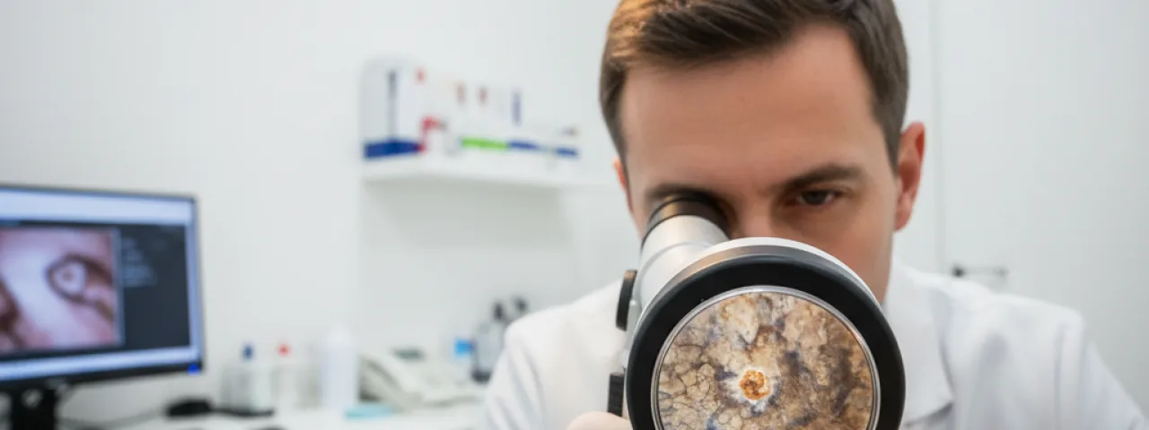
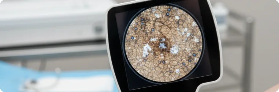
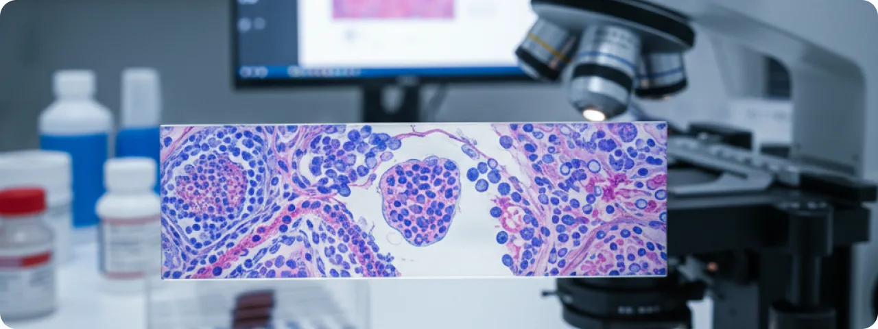
Dermatoscopy is a non-invasive method that magnifies the surface of the skin 10–20 times and improves the accuracy of clinical diagnosis by 25–30%. The doctor analyses pigment structures, vascular patterns and specific features: an atypical pigment network, irregular globules, a blue-white veil. Digital dermatoscopy with Total Body Mapping records every mole at each visit, allowing images to be compared and even minimal changes to be detected. Confocal laser scanning microscopy provides visualisation of skin cells in vivo without a biopsy, at a resolution approaching that of histology. Excisional biopsy remains the gold standard of definitive diagnosis: the suspicious lesion is removed completely with a 1–3 mm margin for morphological examination, with assessment of Breslow thickness, Clark level of invasion and mitotic index. Where necessary, sentinel lymph node biopsy is performed — when the tumour is thicker than 0.8 mm.
Current approaches to treating melanoma
The treatment strategy is determined by the stage of the disease and the molecular and genetic profile of the tumour. Surgical excision is the first-line method in localised disease: the margin is 0.5 cm for melanoma in situ, 1 cm for a thickness of up to 2 mm, and 2 cm above 2 mm. The real breakthrough of the past decade has been immunotherapy with immune checkpoint inhibitors. Anti-PD-1 drugs (pembrolizumab, nivolumab) and anti-CTLA-4 drugs (ipilimumab) have raised five-year survival in metastatic melanoma from 5–10% to 40–50%. The combination of nivolumab and ipilimumab produces a response in up to 58% of patients. Targeted therapy is used where a BRAF V600 mutation is present (found in 40–50% of melanomas): combinations of BRAF and MEK inhibitors (dabrafenib plus trametinib) produce a response in more than 70% of patients. Adjuvant therapy after surgery in stage IIB and above reduces the risk of recurrence by 35–40%. Radiotherapy is used for inoperable cases and for palliative control of metastases.
Prevention and skin self-examination
The most effective strategy against melanoma combines primary prevention with regular monitoring. Limit time in direct sun during peak hours (11:00 to 16:00), apply a sunscreen with SPF 50+ every two hours and after swimming, and choose clothing with UPF protection and a wide-brimmed hat. Give up sunbeds entirely — IARC (the International Agency for Research on Cancer) classifies artificial UV radiation as a group I carcinogen. Carry out a monthly self-examination using the ABCDE rule: use two mirrors to check your back, and do not forget the scalp, the spaces between the fingers and toes, the soles and the area under the nails. Photograph your moles on a smartphone in the same light and at the same scale — this makes changes far easier to track. See a dermatologist for a routine dermatoscopic examination at least once a year. Patients at increased risk should have digital total body mapping and an examination every 3–6 months.
When to seek help urgently
Do not delay seeing a dermato-oncologist if you notice any of the following: a mole growing rapidly over a few weeks; a change in its colour, or uneven colouring appearing; edges becoming indistinct or notched; the mole bleeding, weeping or crusting without any obvious injury; persistent itching, burning or tenderness. Pay particular attention to the "ugly duckling sign" — a mole that looks strikingly different from all the others on your body. New pigmented lesions appearing after the age of 35 also need professional assessment, since new naevi are unusual at that age. Patients with a family history, many atypical naevi or previous skin cancer should be kept under regular follow-up. Remember the main rule: better one unnecessary visit to the doctor than a melanoma missed at an early stage.
Related reading

Did not find the answer to your question?
Every case is individual. If you have noticed changes in your skin, or have symptoms that concern you, book a consultation.