Pre-cancerous skin conditions: what you need to know
What are pre-cancerous skin conditions?
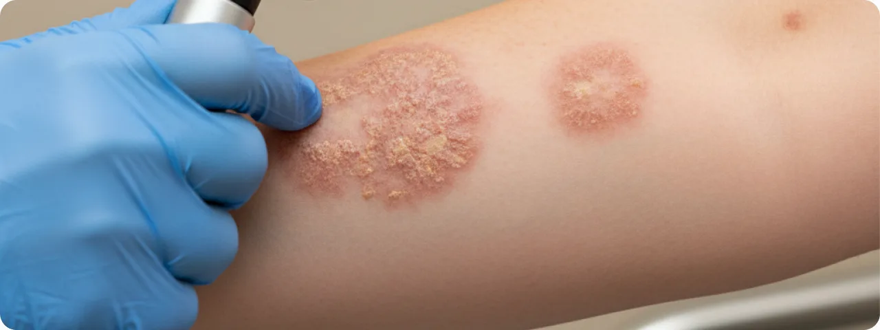
Pre-cancerous skin conditions are pathological changes in the tissue that carry an increased risk of transforming into a malignant tumour. It is important to understand that a pre-cancerous condition is not yet cancer, but without proper attention and treatment it can progress. According to WHO data, up to 60% of cases of squamous cell skin cancer develop from pre-cancerous lesions. The main pre-cancerous skin conditions include: actinic keratosis (the most common — found in 40–60% of fair-skinned people over 40), Bowen's disease (intraepidermal carcinoma in situ), leukoplakia (a lesion of the mucous membranes), keratoacanthoma (a rapidly growing dome-shaped lesion), cutaneous horn (a conical horn-like growth), chronic scars and non-healing ulcers (Marjolin's ulcer), and certain types of naevus — particularly dysplastic (atypical) naevi. The key difference between a pre-cancerous condition and cancer is the absence of invasion: the abnormal cells do not penetrate beyond the basement membrane of the epidermis. That is why detecting and treating pre-cancerous conditions in time is the most effective strategy for preventing skin cancer.
Types of pre-cancerous lesion and how they look
Each type of pre-cancerous condition has characteristic clinical features that help the doctor to reach a provisional diagnosis:
- Actinic keratosis: rough, scaly patches ranging from a few millimetres to 2–3 cm. The colour ranges from skin-coloured to reddish-brown. They appear on exposed skin: the face, ears, scalp (in balding men), the backs of the hands and the forearms. They feel like sandpaper. They may itch or be slightly sore. The risk of a single lesion transforming into squamous cell carcinoma is 5–10% over 10 years, but where there are many lesions the cumulative risk rises considerably.
- Bowen's disease: a sharply demarcated reddish plaque with a scaly or crusted surface, often on the trunk or the lower legs. It enlarges slowly over months and years. It is a carcinoma in situ — the cells are already atypical but confined to the epidermis. Untreated, it progresses to invasive squamous cell carcinoma in 3–5% of cases. It is important to distinguish it from eczema, psoriasis and fungal infection.
- Keratoacanthoma: a rapidly growing dome-shaped lesion with a crater filled with keratin, resembling a volcano. It grows over 2–8 weeks, reaching 1–2 cm. It may regress on its own within 4–6 months, leaving a scar. Because it resembles squamous cell carcinoma both clinically and histologically, surgical removal with histology is recommended.
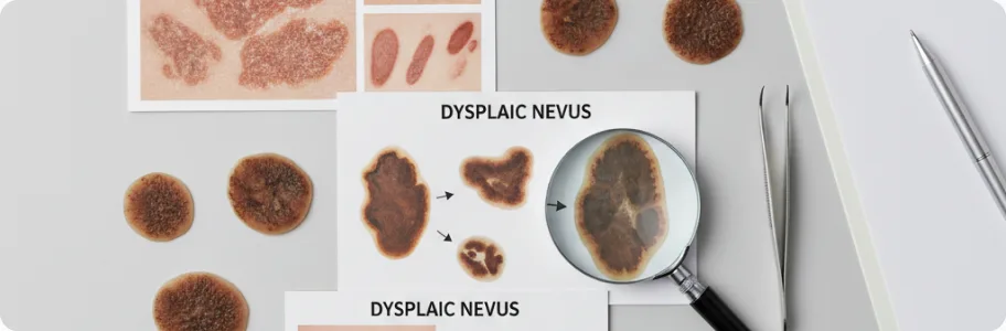
- Dysplastic (atypical) naevus: a mole with an irregular shape, uneven edges and uneven colouring (brown, pink and reddish zones within a single lesion). The diameter is usually more than 5 mm. It may be flat or slightly raised. Dysplastic naevus syndrome (multiple atypical moles plus a family history of melanoma) increases the risk of melanoma 10–15 times. It requires regular monitoring with digital dermatoscopy.
Risk factors: who is at risk
Pre-cancerous skin conditions develop through a combination of external and internal factors. Ultraviolet radiation is the main risk factor: the cumulative dose of UV exposure over a lifetime correlates directly with the likelihood of developing actinic keratosis and squamous cell carcinoma. Sunburn in childhood and adolescence is particularly dangerous — it raises the risk of melanoma by 80%. Skin phototype plays a key role: people with Fitzpatrick phototype I–II (fair skin, fair hair, blue or green eyes, a tendency to burn) have a 5–10 times higher risk than those with phototypes IV–VI. Age — the frequency of pre-cancerous conditions doubles with each decade after 40. Immunosuppression greatly increases the risk: transplant recipients have a 65–250 times higher risk of squamous cell skin cancer. Occupational contact with carcinogens (arsenic, coal tar, ionising radiation), chronic skin inflammation, burn scars and non-healing ulcers are also risk factors. Heredity plays a part in certain genetic syndromes: xeroderma pigmentosum, Gorlin syndrome and dysplastic naevus syndrome.
Diagnosing pre-cancerous conditions
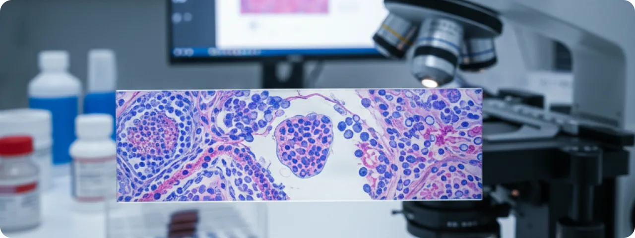
Early diagnosis of pre-cancerous conditions is the basis of effective skin cancer prevention. Clinical examination by a dermatologist includes assessment of every skin lesion using the ABCDE rule: Asymmetry, Border (irregular edges), Color (uneven), Diameter (more than 6 mm) and Evolution (change over time). Dermatoscopy is a non-invasive examination with a dermatoscope that allows skin structures invisible to the naked eye to be seen. The accuracy of melanoma diagnosis with dermatoscopy reaches 90–95%, compared with 65–75% on clinical examination. Digital dermatoscopy with photographic records allows lesions to be followed over time — comparing images at intervals of 3–6 months. Confocal microscopy is a non-invasive method of imaging the skin at cellular level, which in difficult cases helps to avoid an unnecessary biopsy. Biopsy with histology is the gold standard of diagnosis. It is performed where malignant transformation is suspected or where the dermatoscopic picture is ambiguous. It determines the cell type, the degree of atypia and the depth of involvement precisely.
Treatment and prevention
The choice of treatment depends on the type of pre-cancerous condition, its size and site, and the patient's general health. Cryotherapy with liquid nitrogen is the most common treatment for actinic keratosis: quick, effective (up to 95% for isolated lesions) and requiring no anaesthetic. Topical therapy: 5-fluorouracil (Efudex cream) is a cytotoxic drug that selectively destroys atypical cells; imiquimod (Aldara) is an immunomodulator that stimulates a local immune response; ingenol mebutate and diclofenac are also used. Photodynamic therapy (PDT) involves applying a photosensitiser followed by exposure to a special light source; it is effective for large affected areas. Surgical excision is essential for keratoacanthoma and Bowen's disease and provides material for histology. Prevention includes: daily use of sunscreen at SPF 30–50+ even in cloudy weather, wearing protective clothing and hats, avoiding the sun at peak hours (10:00–16:00), giving up sunbeds entirely, an annual dermatological examination with dermatoscopy, and a monthly self-examination of the skin recording any changes. Remember: a pre-cancerous condition is not a sentence but a signal to act. Detected and treated in time, the outlook is entirely favourable.
Common questions about pre-cancerous skin conditions
Does a pre-cancerous condition always turn into cancer? — No, far from always. The risk of transformation depends on the type of lesion: for a single actinic keratosis it is 5–10% over 10 years, and for Bowen's disease 3–5%. But it is precisely the unpredictability of the process that makes treatment essential. Can I identify a pre-cancerous condition myself? — Self-examination of the skin is an important tool for early detection. Look out for new lesions, changes in existing moles, patches that do not heal and rough areas of skin. The final diagnosis, however, is made only by a doctor after dermatoscopy and, where necessary, a biopsy. Do all suspicious moles need to be removed? — Not necessarily. Dysplastic naevi that show no change can be monitored with digital dermatoscopy. Removal is indicated where malignant transformation is suspected or where monitoring shows marked change. How often should I see a dermatologist? — For the general population, an annual examination is recommended. Where risk factors are present (multiple atypical naevi, a family history of melanoma, previous skin cancer, immunosuppression) — every 3–6 months. Does diet affect the risk of skin cancer? — Research suggests that antioxidants (vitamins C and E, beta-carotene), omega-3 fatty acids and green tea may have a modest protective effect, but no food replaces sun protection and regular examinations.
Related reading

Did not find the answer to your question?
Every case is individual. If you have noticed changes in your skin, or have symptoms that concern you, book a consultation.