Couperose: why it appears and how it is treated

What is couperose and why does it appear?
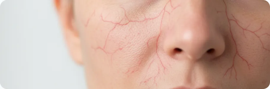
Couperose is a chronic skin condition characterised by persistent dilation of the small blood vessels (capillaries, arterioles and venules) in the superficial layers of the dermis. The visible networks of vessels — telangiectasias — appear most often on the cheeks, the sides of the nose, the chin and the forehead. Couperose is not merely a cosmetic problem: it indicates impaired microcirculation and weakness of the vessel wall. The main causes include a genetic predisposition (weakness of the connective tissue of the vessels), hormonal changes (pregnancy, the menopause, oral contraceptives), chronic exposure to ultraviolet light, abrupt changes in temperature, excessive alcohol and spicy food, and prolonged use of corticosteroids.
Stages and symptoms of couperose
Couperose develops gradually, through several stages. Early diagnosis substantially improves the outlook for treatment:
- stage I (erythrosis): intermittent flushing of the facial skin in response to triggers — cold, heat, hot drinks, stress. The redness lasts from a few minutes to a few hours and is accompanied by a sensation of flushing and mild tingling. At this stage the vessels can still return to normal tone, so the outward signs resolve on their own.
- stage II (telangiectasia): persistent, visible red or bluish spider veins and networks appear on the skin. They no longer disappear once the provoking factor is removed. The skin becomes more sensitive, and mild flaking and dryness may appear. Cosmetics begin to cause irritation. This is the stage at which patients most often come to a dermatologist.
- stage III (rosacea-like couperose): areas of persistent congestive redness form, and the network of vessels becomes denser and more noticeable. Inflammatory lesions — papules — may join in. The skin takes on an uneven tone and feels coarser. Tissue nutrition is impaired, which leads to premature ageing of the skin in the affected areas.
- stage IV (extensive couperose): the vascular changes cover a large area of the face and the skin takes on a permanent bluish-red tone. The skin may thicken and nodules may form. At this stage treatment requires a combined approach using procedural methods.
Instrumental diagnosis of couperose
Diagnosis rests on visual examination and instrumental methods. Dermatoscopy allows the superficial vessels to be assessed under magnification, the type of telangiectasia to be determined (linear, arborising, stellate, punctate) and the depth of involvement to be judged. Capillaroscopy is a method of assessing microcirculation that allows individual capillaries to be visualised and the speed of blood flow to be assessed. Laser Doppler flowmetry measures the intensity of microcirculation in real time. Ultrasound of the skin allows the thickness of the dermis and the state of the deeper vessels to be assessed. Photographic documentation under standardised lighting (polarised, UV) helps to track progress and the effectiveness of treatment. It is also important to obtain a full blood count and a coagulation profile and to assess hormonal status, in order to identify systemic causes.
Effective treatments for couperose
Treatment should be comprehensive and include both procedures and medical support. Laser therapy (Nd:YAG 1064 nm, diode 940 nm) is the gold standard for removing telangiectasias: the laser beam selectively heats the haemoglobin in the vessel and causes it to coagulate; 2–5 sessions are needed at intervals of 3–4 weeks. IPL (intense pulsed light) therapy is effective for diffuse redness and fine vessels; a course of 3–6 treatments. Electrocoagulation is used for isolated larger vessels. Microsclerotherapy involves injecting a sclerosing solution into the dilated vessel. Topical treatments: a cream with azelaic acid (15–20%) reduces erythema, products with vitamin K and rutin strengthen the vessel wall, and brimonidine (0.33%) is a topical alpha-adrenergic agonist for rapidly reducing redness. Mesotherapy with vitamin C, peptides and hyaluronic acid improves microcirculation and strengthens the vessels.
Skin care and prevention
Morning routine: gentle cleansing with a milk or micellar water (free of sulphates, alcohol and abrasive particles), a serum with niacinamide (vitamin B3, 4–5%) to strengthen the vessel wall, a moisturiser with ceramides and panthenol to repair the barrier, and sun protection at SPF 50+ with mineral filters (zinc oxide, titanium dioxide) — UV is one of the main triggers of couperose. Evening routine: delicate cleansing with a cleansing oil, an alcohol-free toner with horse chestnut or ginkgo biloba extract, a serum with vitamin C (ascorbyl glucoside — a stable form that does not irritate), and a night cream with retinaldehyde (a gentle retinoid) to stimulate renewal. General rules: avoid abrupt changes in temperature, do not use saunas or steam baths, limit alcohol and spicy food, and use thermal water to calm the skin during the day.
When to see a dermatologist
See a dermatologist if you notice persistent facial redness that does not fade within a few hours, if visible spider veins or networks have appeared on the skin, or if your skin has become excessively sensitive to cosmetics and to changes in temperature. An immediate consultation is needed where progression is rapid — new telangiectasias appearing week by week — where inflammatory lesions (papules, pustules) have joined in, or where new areas of the face are becoming involved. See a doctor as well if home care produces no result within 4–6 weeks, if the couperose appeared after starting new medication (corticosteroids, vasodilators), or if the visible vascular changes are affecting your quality of life and self-esteem.
Related reading


Did not find the answer to your question?
Every case is individual. If you have noticed changes in your skin, or have symptoms that concern you, book a consultation.