Onycholysis: why the nail separates and how it is restored
What is onycholysis and why does it occur?
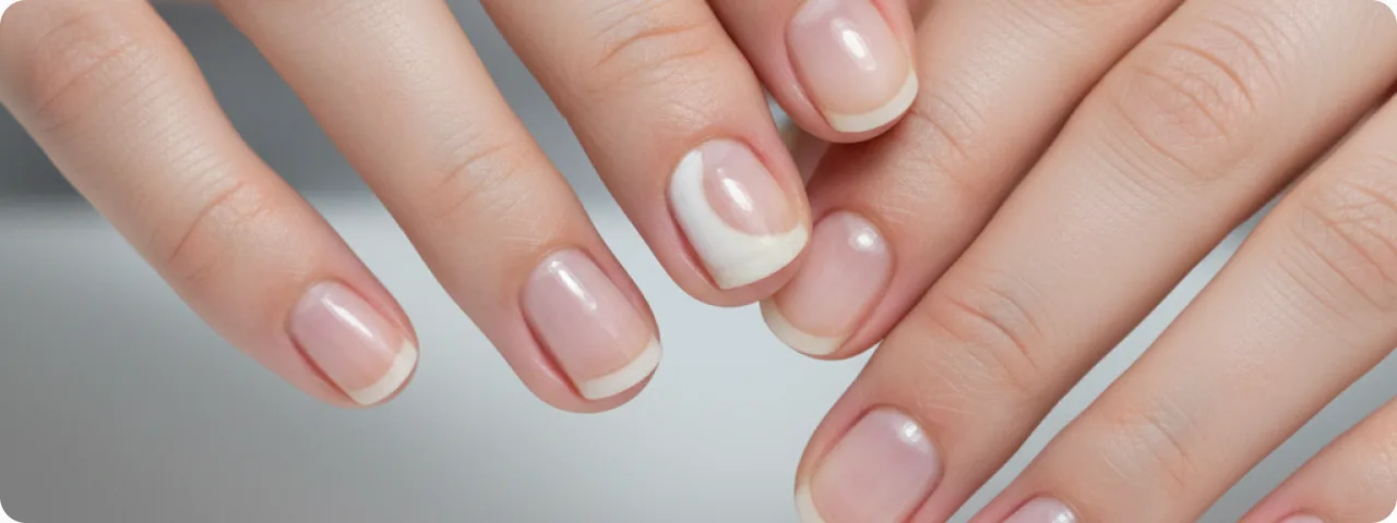
Onycholysis is separation of the nail plate from the nail bed, usually beginning at the free edge of the nail and gradually advancing towards the matrix. It affects 2–5% of the population, with women affected 2–3 times as often as men. The causes are varied: traumatic (mechanical damage, tight shoes, aggressive manicure), infectious (fungal — up to 30% of cases; bacterial — particularly Pseudomonas aeruginosa), dermatological (psoriasis, eczema, lichen planus), systemic (thyroid disease, anaemia, diabetes), drug-related (tetracyclines, fluoroquinolones, chemotherapy) and allergic (a contact reaction to gel polishes, acrylic materials or household chemicals). In 30% of cases the exact cause cannot be established — such onycholysis is called idiopathic.
Signs and stages of onycholysis
Onycholysis develops gradually and has characteristic clinical features, which differ according to the stage and the cause of separation:
- Early stage (partial separation): the nail begins to lift from the bed at the distal or lateral edge. An air space forms beneath the separated area, giving the nail a whitish or yellowish tone. The nail keeps its normal shape and thickness, but a cavity can be felt when pressure is applied. The separated area usually does not exceed 30% of the nail plate. At this stage the process is often reversible once the cause is removed.
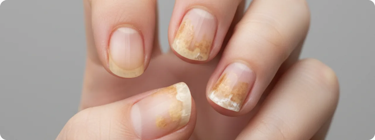
- Progressive stage (extensive separation): the separated area increases to 50–70% of the nail plate. Hyperkeratotic material, dirt and microorganisms may accumulate beneath the nail. The colour of the separated area changes: a green tone indicates Pseudomonas, yellow-brown indicates a fungal infection, and greyish-white a traumatic cause. An unpleasant smell develops, particularly where there is secondary infection.
- Total onycholysis (complete separation): the nail plate separates completely from the bed and may fall off spontaneously (onychomadesis). The nail bed becomes thickened, dry and covered with hyperkeratosis. The risk of secondary infection is at its highest. Full recovery at this stage takes 6–12 months on the hands and 12–18 months on the feet. It requires comprehensive systemic treatment and the underlying cause must be established.
- Associated changes in the nail plate: thickening or thinning of the nail, longitudinal and transverse ridges, changes in colour (leukonychia, melanonychia) and brittleness of the free edge. In psoriatic onycholysis, "oil drop" patches — yellow-brown areas beneath the nail — are characteristic. In fungal disease there is subungual hyperkeratosis with a yellowish colour. In bacterial infection the colour is greenish or black with a strong smell.
Diagnosis: establishing the cause
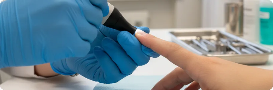
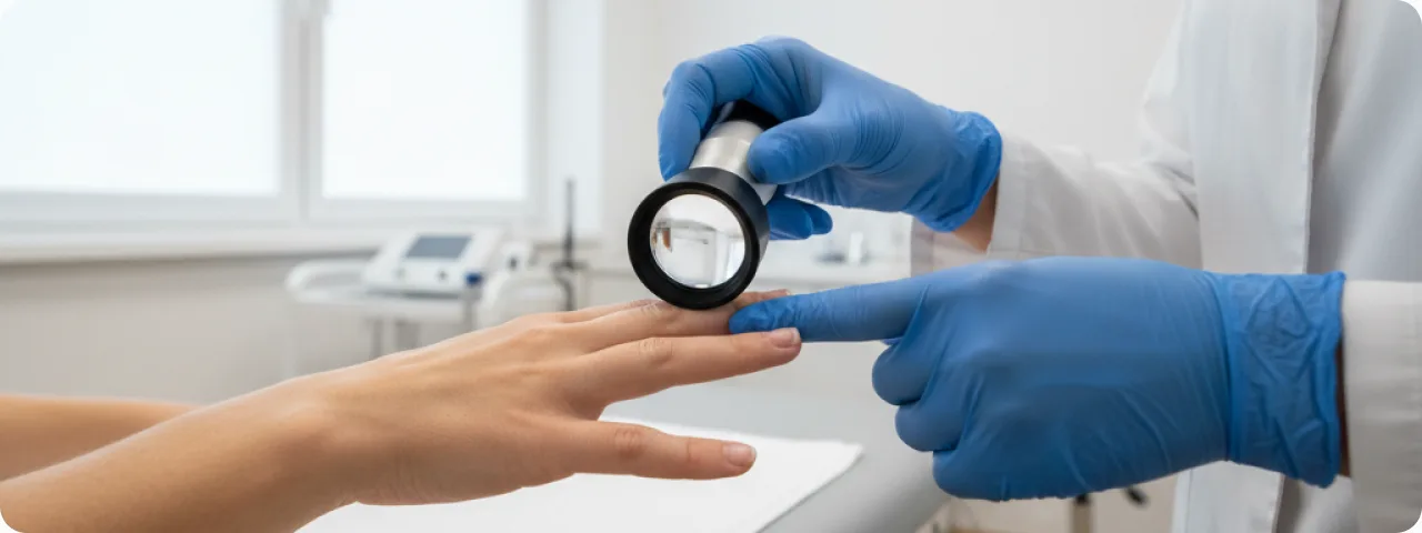
Correct diagnosis is the key to effective treatment, because the therapy depends entirely on the cause. Microscopy and the KOH test are essential to exclude or confirm a fungal infection. The result is positive in 30–50% of cases of onycholysis. Bacterial culture is done where a bacterial infection is suspected, particularly where the colour beneath the nail is green-black (Pseudomonas aeruginosa). Dermatoscopy of the nail is a non-invasive way of assessing the capillaries of the nail bed and identifying splinter haemorrhages, oil-drop patches and other pathognomonic features. Blood tests include a full blood count, a thyroid panel (TSH, T3, T4), iron and ferritin, and blood glucose — to exclude systemic causes. Histology of a nail biopsy is used in difficult cases, where non-invasive methods give no clear answer. Allergy testing (patch tests) is used where contact dermatitis from cosmetic materials or household chemicals is suspected.
Current treatment of onycholysis
Treatment depends on the cause and the extent of involvement. Removing the provoking factor is the first and most important step: stopping contact with the allergen, changing footwear, giving up aggressive cosmetic procedures, and changing a drug that is responsible. Topical antifungal therapy (in fungal onycholysis): amorolfine 5% lacquer — applied 1–2 times a week for 6–12 months; ciclopirox 8% lacquer — applied daily. Systemic antifungal therapy: terbinafine 250 mg/day (3 months for the hands, 6 months for the feet) is the drug of choice; itraconazole as pulse therapy. For bacterial infection: topical antibiotics (gentamicin, ciprofloxacin) and antiseptic soaks with chlorhexidine or povidone-iodine. In psoriatic onycholysis: topical corticosteroids and calcipotriol, triamcinolone injections into the nail matrix, and systemic therapy with methotrexate or biologic agents. Restorative treatment: biotin (5–10 mg/day), zinc and iron preparations, and oils to strengthen the nails (jojoba, almond, castor).
Prevention and nail care
Prevention rests on proper nail care and avoiding factors that cause injury. Cut the nails short and straight, without rounding the corners — long nails are more vulnerable to mechanical damage and separation. Choose a gentle manicure and pedicure: do not cut the cuticle deeply, avoid metal pushers under the nail plate, and do not remove a coating by peeling it off — that damages the upper layer of the nail. Wear protective gloves when handling household chemicals, solvents and detergents — chemical exposure is one of the most frequent causes of onycholysis in women. Choose good-quality gel polishes and extension materials — cheap products often contain harsh ingredients that cause contact allergy and separation. Dry the nails thoroughly after contact with water — a damp environment encourages fungal and bacterial infection. Wear comfortable shoes with enough room for the toes — tight shoes exert constant pressure on the toenails. Eat a balanced diet: enough biotin, zinc, iron and B vitamins is necessary for healthy nail growth.
When to see a dermatologist
See a dermatologist if you notice that a nail has begun to separate from the bed — even minor separation can progress without proper treatment. An immediate consultation is needed if the colour beneath the nail changes (green, black or brown), if pain or swelling appears around the nail, or if there is an unpleasant smell from under the nail plate — these are signs of secondary infection requiring specific treatment. See a specialist if onycholysis has developed without an obvious cause (no injury and no contact with chemicals) — this may indicate systemic disease (thyroid disorder, anaemia, diabetes). A consultation is also essential if onycholysis recurs after treatment, if several nails are affected at once, or if you have noticed associated skin changes (a rash, redness, itching), which may point to a dermatological disease.
Related reading
Did not find the answer to your question?
Every case is individual. If you have noticed changes in your skin, or have symptoms that concern you, book a consultation.