Pigmented nail lesions: benign versus malignant

Longitudinal melanonychia: what is it?
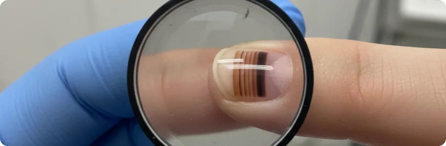
Longitudinal melanonychia is a pigmented band running from the nail matrix to the free edge of the nail. It may be an entirely benign finding or the first sign of melanoma. In people with darker skin phototypes (IV–VI) melanonychia is found in 77% of cases and is usually physiological — so-called racial or ethnic melanonychia. In fair-skinned people it is less common and more often requires detailed assessment. Benign causes include activation of melanocytes by injury, friction or pregnancy, medication (antimalarials, cytotoxics, zidovudine), fungal infection, or a naevus of the nail matrix. The malignant cause — subungual melanoma — accounts for less than 3% of all cases of melanonychia, but it is what makes the differential diagnosis clinically important.
Benign pigmented lesions of the nails
Most pigmented nail changes are benign. Understanding their features helps to avoid unnecessary anxiety while not missing the dangerous signs.
- Melanocytic naevus of the matrix: the most common cause of longitudinal melanonychia in children and adolescents. The band has a uniform colour (from light to dark brown), clear, even borders and a stable width over years. Dermatoscopy shows regular parallel lines of the same thickness and spacing. The naevus may enlarge slowly while the child is growing, which is normal.
- Subungual haematoma: a dark patch beneath the nail after injury (crushing, a knock). The key difference from melanoma is that a haematoma migrates distally as the nail grows and disappears completely within 6–9 months. It does not form a longitudinal band from the cuticle. On dermatoscopy it has characteristic rounded globular structures. If the patient does not remember an injury, the diagnosis needs confirmation.
- Drug-induced melanonychia: develops with certain medications — zidovudine (in 40% of patients with HIV), hydroxyurea, cyclophosphamide, doxorubicin and antimalarials. Several nails are usually affected at once, which distinguishes it from melanoma. After the drug is stopped, the pigmentation gradually fades over several months.
- Fungal melanonychia and matrix lentigo: some dermatophytes and moulds (Trichophyton rubrum var. nigricans, Scytalidium dimidiatum) produce a dark pigment that mimics melanonychia. It is diagnosed by microscopy and culture. Lentigo of the nail matrix is focal hyperplasia of melanocytes without atypia, characteristic of middle-aged and older people. Both conditions must be distinguished from melanoma by dermatoscopy and, in doubtful cases, by biopsy.
Malignant lesions: subungual melanoma
Subungual melanoma is a rare but the most dangerous pigmented lesion of the nail, accounting for 1–3% of all melanomas in fair-skinned people and up to 15–35% in people with darker skin. The key warning signs follow the ABCDEF rule: Age (over 50), Band (wider than 3 mm with uneven colouring), Change (progression over weeks or months), Digit (involvement of the thumb or great toe), Extension (spread of pigment onto the nail fold — Hutchinson's sign) and Family history (melanoma in relatives). Dermatoscopy reveals irregular lines of varying thickness, micro-Hutchinson's sign and interrupted pigment structures. The diagnosis is confirmed only by biopsy of the matrix with histopathology and immunohistochemistry (S-100, HMB-45, Melan-A). The average interval between the first symptom and the diagnosis is 2–3 years, which explains the late stage at detection and the poorer five-year survival.
The differential diagnosis, step by step
Clinical assessment begins with the history: the age at which the band appeared, how it has changed, how many nails are affected, medication, injuries and the family history. Dermatoscopy is the first instrumental method — a regular pattern of parallel lines of the same colour and thickness favours a benign lesion, whereas irregular lines, several colours and blurred borders point to malignancy. In doubtful cases, follow-up with photographic documentation every 3–6 months is recommended. Indications for biopsy: a new band in a fair-skinned adult, a width over 3 mm, progressive widening, several colours, Hutchinson's sign, or dermatoscopic irregularity. Biopsy of the matrix is the gold standard, using a punch, incisional or excisional technique depending on where the pigment lies. The histological report determines what follows: observation, repeat biopsy or surgery.
Management: from observation to surgery
Management depends on the outcome of the differential diagnosis. Benign lesions — ethnic melanonychia, a stable naevus, drug-induced pigmentation — need only regular observation with photographs. A subungual haematoma resolves on its own; where the diagnosis is in doubt, dermatoscopy is repeated after 1–2 months to confirm distal migration. A naevus of the nail matrix may be removed at the patient's request or if it changes suspiciously. For subungual melanoma in situ, a wide excision is performed with complete removal of the nail apparatus and preservation of the digit. Invasive melanoma requires amputation of the distal phalanx; where the Breslow thickness exceeds 1 mm, sentinel lymph node biopsy is recommended. Advanced stages are treated according to systemic therapy protocols — immunotherapy (nivolumab, pembrolizumab) and targeted therapy where BRAF mutations are present.
Advice for patients and self-monitoring
Regular self-examination of all 20 nails is the basis of early detection. Examine the nails in good light at least once a month, looking for new longitudinal bands, changes in the colour or width of existing bands, and destruction of the nail plate without an obvious cause. Photograph any suspicious area for comparison over time — even minimal change over 3–6 months can be clinically significant. Do not hide dark bands under polish or a gel coating — that delays diagnosis. Remember: a single stable band of uniform colour up to 3 mm wide that has not changed for years is most likely benign. A new band after the age of 50, rapid widening, several colours or spread of pigment beyond the nail, however, call for a prompt dermatological consultation. People with a family history of melanoma, multiple naevi or immunosuppression should have their nails examined by a specialist every year.
Related reading


Did not find the answer to your question?
Every case is individual. If you have noticed changes in your skin, or have symptoms that concern you, book a consultation.